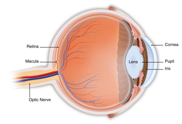
Retina
Understanding the Retina
The retina is a thin layer of nerve tissue that lines the inner wall of the eye. This layer of the eye is the light sensitive layer that is activated when light from the front of the eye is focused on to it. The focused light activates electrical signals in the retina that then are transmitted via the optic nerve to the brain where this information is converted into an image. The retina functions in much the same way as film in a camera. There are many conditions that affect the retina that can lead to temporary or permanent vision loss.
What is a retina specialist?
A retina specialist is an ophthalmologist (Eye MD) who has completed an additional 2 year training fellowship in medical and surgical treatment of retinal disease.
What testing is done to evaluate the retina?
When you see an ophthalmologist for an evaluation of a retinal condition your eyes will be dilated with special dilating eye drops to improve the view to the back of the eye. The ophthalmologist will use a diagnostic microscope called a slit lamp and special lenses to view the retina. The peripheral retina may be examined with a head mounted ophthalmoscope called an indirect ophthalmoscope. Various diagnostic test may be performed which may include ultrasound of the eye, photographs, optical coherence tomography (OCT) or fluorescein angiography of the retina.

Testing for Retinal Disease
Common Diseases of the Retina
Diabetic Retinopathy
Posterior Vitreous Detachment (PVD)
The pulling or tugging of the retina in these areas activates the retina producing a flash of light. Patient’s experiencing a PVD may see these flashes that look like a camera flash or lightening bolt in their peripheral vision. Also, as the vitreous releases from the back of the eye the edges of the vitreous can be seen as new floaters. It is important to alert your ophthalmologist if you experience these symptoms. Rarely, the retinal traction from a PVD may result in a tear of the retina or retinal detachment.
Retinal Detachment
Macular Edema
Lattice Degeneration
Macular Degeneration
Retinal Tear
Epiretinal Membrane (ERM)
Retinal Vein Occlusion
Macular Hole
